Review sheet exercise 30 anatomy of the heart – Embarking on a journey through the intricate anatomy of the heart, Review Sheet Exercise 30 unveils the secrets of this vital organ. Prepare to navigate the heart’s structure, its role in the circulatory system, and the nuances of its internal workings.
Unraveling the heart’s external features, we explore the protective layers of the pericardium and the significance of the coronary sulcus. The atrioventricular sulcus, a crucial boundary, guides us into the heart’s interior.
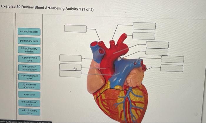
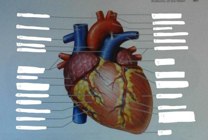
Anatomy of the Heart: Review Sheet Exercise 30 Anatomy Of The Heart

The heart, located in the mediastinum, is a muscular organ approximately the size of a clenched fist. It serves as the central pump of the circulatory system, responsible for propelling blood throughout the body to deliver oxygen and nutrients while removing waste products.
External Heart Anatomy
The heart is enclosed within a double-layered sac called the pericardium. The outer layer, the fibrous pericardium, provides structural support, while the inner layer, the serous pericardium, secretes a lubricating fluid to reduce friction. The coronary sulcus, a groove on the surface of the heart, marks the boundary between the atria and ventricles.
The atrioventricular sulcus, located inferior to the coronary sulcus, separates the atria from the ventricles and accommodates the atrioventricular valves.
Internal Heart Anatomy
Chambers of the Heart
The heart comprises four chambers: two atria (left and right) and two ventricles (left and right).
| Chamber | Location | Function | Blood Flow Direction |
|---|---|---|---|
| Right Atrium | Superior to right ventricle | Receives deoxygenated blood from the body | From superior and inferior vena cava to right ventricle |
| Left Atrium | Superior to left ventricle | Receives oxygenated blood from the lungs | From pulmonary veins to left ventricle |
| Right Ventricle | Inferior to right atrium | Pumps deoxygenated blood to the lungs | From right atrium to pulmonary artery |
| Left Ventricle | Inferior to left atrium | Pumps oxygenated blood to the body | From left atrium to aorta |
Valves of the Heart
The heart valves prevent backflow of blood and maintain unidirectional blood flow.
| Valve | Location | Structure | Function | Clinical Significance |
|---|---|---|---|---|
| Tricuspid Valve | Between right atrium and right ventricle | Three cusps (leaflets) | Prevents backflow from right ventricle to right atrium | Tricuspid regurgitation |
| Pulmonary Valve | Between right ventricle and pulmonary artery | Three cusps | Prevents backflow from pulmonary artery to right ventricle | Pulmonary stenosis |
| Mitral Valve (Bicuspid Valve) | Between left atrium and left ventricle | Two cusps | Prevents backflow from left ventricle to left atrium | Mitral regurgitation, mitral stenosis |
| Aortic Valve | Between left ventricle and aorta | Three cusps | Prevents backflow from aorta to left ventricle | Aortic regurgitation, aortic stenosis |
Blood Flow through the Heart
Blood enters the right atrium from the superior and inferior vena cava. It passes through the tricuspid valve into the right ventricle, which pumps it to the lungs via the pulmonary artery. In the lungs, blood releases carbon dioxide and absorbs oxygen, becoming oxygenated.
The oxygenated blood returns to the heart through the pulmonary veins and enters the left atrium. It then flows through the mitral valve into the left ventricle, which pumps it to the body through the aorta.The coronary arteries, which branch off the aorta, supply oxygenated blood to the heart muscle.
Deoxygenated blood from the heart muscle is collected by the coronary veins and drains into the right atrium.The electrical conduction system of the heart coordinates the heart’s contractions. The sinoatrial node (SA node) in the right atrium initiates the electrical impulse, which travels through the atrioventricular node (AV node) and then along the bundle of His to the left and right bundle branches.
These branches distribute the impulse to the ventricular muscle, causing it to contract.
Variations and Anomalies, Review sheet exercise 30 anatomy of the heart
Variations in heart anatomy are common. An atrial septal defect (ASD) is a hole in the wall between the atria, allowing blood to flow between them. Ventricular septal defects (VSDs) are holes in the wall between the ventricles, causing oxygenated and deoxygenated blood to mix.
These variations can lead to various clinical implications, such as murmurs, arrhythmias, and heart failure.
Imaging Techniques
Echocardiography, a non-invasive ultrasound technique, provides detailed images of the heart’s structure and function. It can detect abnormalities such as valve defects, septal defects, and pericardial effusions. Cardiac magnetic resonance imaging (MRI) uses magnetic fields and radio waves to produce high-resolution images of the heart.
It is particularly useful for assessing myocardial perfusion, identifying cardiac tumors, and evaluating congenital heart defects.
Commonly Asked Questions
What is the purpose of the pericardium?
The pericardium provides protection, lubrication, and stability to the heart.
What is the function of the atrioventricular sulcus?
The atrioventricular sulcus separates the atria from the ventricles and provides a pathway for blood flow.
What is the significance of the coronary arteries?
The coronary arteries supply oxygenated blood to the heart muscle itself.